Did you know that some insects are being x-rayed so precisely that their insides show up like tiny alien landscapes?
In the blog entitled “Insect Anatomy Under the X-Ray: Bugs Tell Big Ecological Tales” we explore how cutting-edge imaging using powerful synchrotron X-rays is tearing open the secrets hidden in even the smallest of creatures. What we see inside these bugs isn’t just fascinating — it could reshape how we understand their roles in climate, ecosystems, and even agriculture.
Why Shrinking, Opaque Things Matter
Most of us think insects as small, simple creatures — but appearances deceive. Under their hard shells or translucent wings lie complex anatomy: respiratory tubes, digestive tracts, muscles, feeding structures. Traditional methods like dissection or light/electron microscopy have helped scientists peek inside. Yet those methods often destroy the specimen, or only allow a partial view.
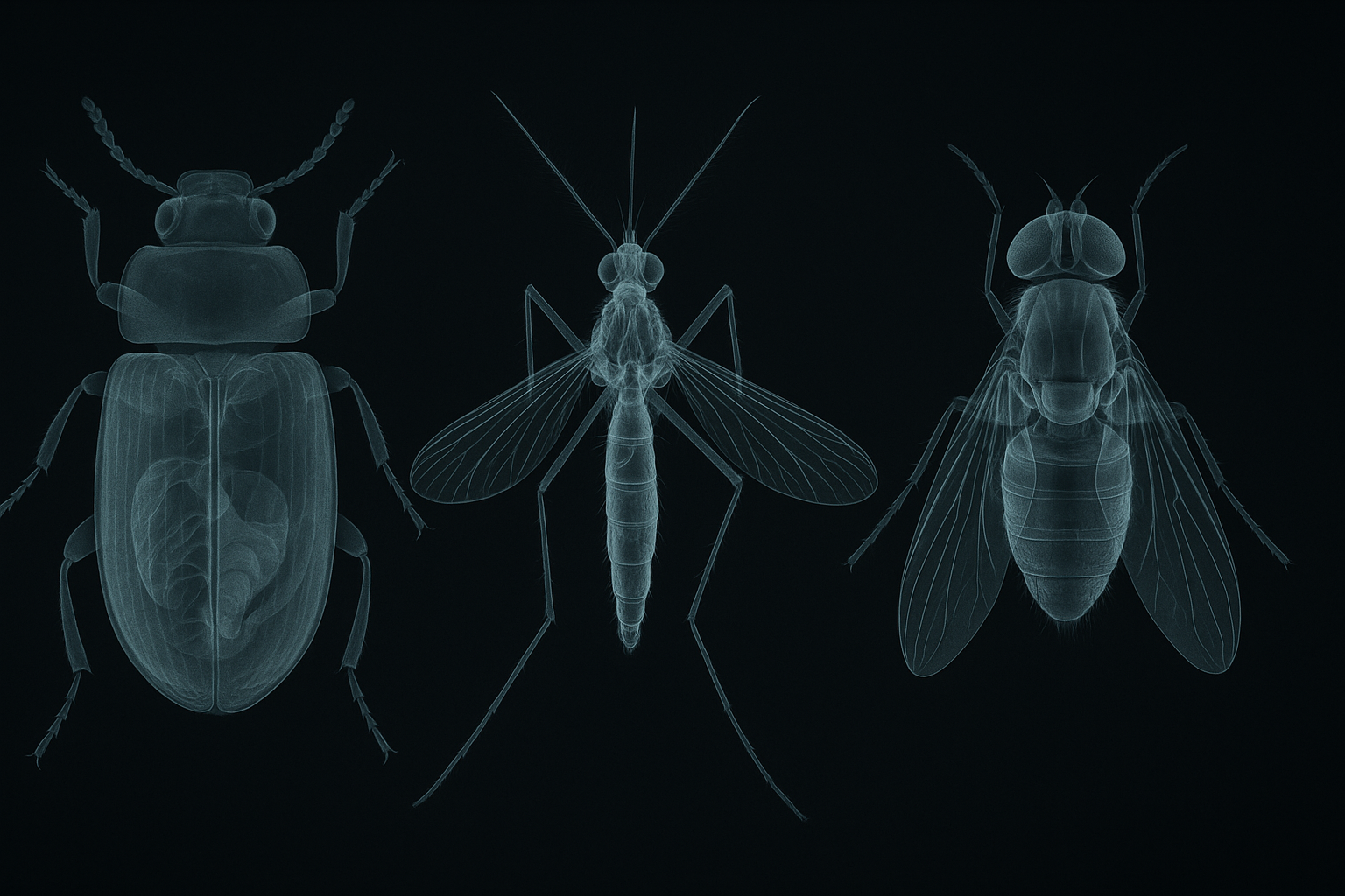
Enter synchrotron X-ray imaging — a technology born for physics labs, now retooled for biology. With synchrotron-based micro-computed tomography (SR-µCT) and phase-contrast X-ray imaging, researchers can peer inside insect bodies in three dimensions, without slicing or staining, often at resolutions down to micrometres. The result: intact, fully detailed anatomical maps.
In simple words — think of it as a full-body “MRI for bugs” — except often much sharper and able to capture soft tissue and even internal air-filled tracheal tubes that normal X-rays or standard microscopes miss.
What Has the X-Ray Revealed — Some Examples
The breathing bugs
One of the earliest triumphs came when scientists used synchrotron X-rays to watch how opaque insects breathe. Using phase-contrast imaging, they directly observed the rhythmic compression of tiny tracheal tubes — some as narrow as 10 micrometres — which carry oxygen through the insect’s body.
This breathing mechanism had been largely inferred in translucent or large insects; now, even common beetles with dark, thick exoskeletons reveal the mechanics of respiration in full. The implication: even small changes in climate — temperature, humidity, oxygen levels — could impact how insects breathe. Maybe some insects already struggle quietly, inside, as heat or pollution changes their environment.
Hidden architects under the shell
A 2022 study used SR-µCT to describe, in unprecedented detail, the internal organs of a common beetle species — the red flour beetle (Tribolium castaneum) — a pervasive pest in stored grains.
Without cutting the beetle open, researchers mapped its digestive system, internal organs, and exoskeletal internal supports. For agriculture and food-storage sectors (especially in countries like ours, where grains are often stored in rustic conditions), knowing exactly how pests eat, breathe and reproduce can be a game changer.
The tiny, fatal mouth of pests
In a 2024 study, scientists used synchrotron X-ray tomography to investigate a notorious rice pest — the Nilaparvata lugens (Brown Planthopper). They discovered in high resolution the structure of its mouthparts, foregut and the channels used to suck sap from rice plants.
Why does that matter beyond academic curiosity? Because the mouthpart anatomy directly shapes how effectively the insect feeds — and how efficiently it spreads diseases from plant to plant. This knowledge might help design better pest-control strategies, or even discourage pesticide use if biology-based methods (e.g. blocking feeding channels) are used.
Why non-destructive, 3D imaging matters for ecology
Unlike dissection or destructive imaging (which kill or alter the specimen), synchrotron imaging preserves entire insect bodies. This is especially valuable when dealing with rare species, museum specimens, or valuable collectors’ samples (for example, insects trapped in amber, or endangered species).
It also means scientists can share digital 3D models across labs — enabling researchers worldwide to virtually examine the same individual without risk of damage.
Bigger Implications: From Micro-Anatomy to Macro-Ecology
So why should you care — even if you’ve never held a beetle or studied a rice plant? Because insect anatomy, when revealed in 3D and high-resolution, has ripple effects across ecology, climate science, agriculture and biodiversity.
Climate change and insect physiology:
As global temperatures and humidity fluctuate, insect metabolic and respiratory systems — tuned to narrow environmental ranges — may get stressed. Detailed anatomy from synchrotron imaging helps model which species are vulnerable, how heat or drought may harm them, and how their survival or death may ripple through food webs.
Pest control re-imagined:
For pests like the brown planthopper or red flour beetle, understanding exactly how they feed or reproduce could lead to more targeted controls — maybe blocking mouthparts, or using biocontrol agents — reducing reliance on chemical pesticides, which harm beneficial insects and pollinators.
Conservation and biodiversity mapping:
Many insect species remain unknown or under-described. Non-destructive 3D imaging means taxonomists and conservation biologists can catalogue internal features, differentiate species, and preserve digital records — helping biodiversity inventories without killing thousands of fragile specimens.
Research democratization:
With virtual 3D models, even scientists far from synchrotron labs can study insect anatomy. This could catalyse discoveries from small labs in developing countries — a powerful equalizer in global ecological research.
Why This Feels Like a Small Revolution
We tend to think “big science” — telescopes, genetic sequencing, satellites — when we consider climate or biodiversity research. We forget that some of the most dramatic stories play out at scales too small to see with naked eyes.
Watching a beetle breathe. Watching how a planthopper’s mouth is built to pierce living tissue. Watching, somehow, the invisible mechanics of life inside an insect. That is intimate. It’s personal.
Maybe what we call “nature’s small actors” — bugs, beetles, planthoppers, ants — are really the earth’s subtle engineers, quietly shaping ecosystems. And maybe, by giving them X-rays and respect, we start to treat them as more than nuisances — but as indicators, early-warning signals of climate stress, environmental imbalance, ecological loss.
As I see it, synchrotron imaging doesn’t just make pretty pictures: it inaugurates a new era of ecological empathy — for the small, for the overlooked.
Conclusion
The tiny creatures buzzing around our homes, fields, and granaries carry worlds within them — tracheal labyrinths, digestive corridors, feeding needles — all shaped by millions of years of evolution. Through synchrotron X-ray imaging, we are finally peeking in. And what we see matters. It may hold clues about how insects survive (or fail) under changing climates, how pests might be controlled in smarter, gentler ways, how biodiversity can be conserved in digital as well as natural form.
If we care about the future of ecosystems — and whether our crops survive, pollinators thrive, and ecological balance endures — then maybe we need to start paying attention not just to big beasts, but to the tiny bodies we often crush unconsciously underfoot. Maybe, in their bones and tracheae, lies a message: small beings, big stories.
So next time you swat a mosquito, squash a beetle, or brush away a fly — maybe pause. You might be disrupting a little universe. And maybe something inside that insect was carrying subtle wisdom about nature, climate, and life itself.
Author’s Note
I write this after a quiet evening of listening to crickets outside my home — wondering what mysteries those tiny chirpers carry beyond their songs. This piece is not about glorifying bugs, nor about painting them as villains or heroes. It is about paying attention. Because sometimes the smallest bodies store the most important stories. If shining an X-ray light on them makes us pause — and maybe act differently — that, to me, is enough reason to write.
G.C., Ecosociosphere contributor.
References and Further Reading
- Insect imaging at the ANKA synchrotron radiation facility (overview of synchrotron-based insect studies)
- Anatomical study of the red flour beetle with synchrotron X-rays
- Synchrotron-based micro-X-ray computed tomography for insect morphology
- High-resolution 3D imaging of insects with synchrotron X-rays (2022)
- Synchrotron radiation X-ray tomography revealing foregut and mouthpart of brown planthopper (2024)